Übersetzt aus dem Englischen:
Die Indikationen für eine Operation am Deltaband können im Hinblick auf die akute Verletzungssituation betrachtet werden, wobei es eine gewisse Kontroverse über die Notwendigkeit der Reparatur des Deltabandes gibt, und die chronische Situation bei Vorliegen einer langjährigen planovalgus Deformität und/oder medialen Sprunggelenkinstabilität, bei der es wenig Kontroverse über die Reparatur der Struktur gibt. Diese werden im Indikationsabschnitt genauer besprochen. Das Deltaband besteht aus oberflächlichen und tiefen Komponenten, die miteinander verschmelzen. Der oberflächliche Teil des Deltabands ist die Hauptbegrenzung für Valgus im Sprunggelenk und Rückfuß, und die tiefe Komponente widersteht der externen Rotation. Anatomisch verläuft das oberflächliche Deltaband als fächerförmige Struktur vom medialen Malleolus, wobei die beschriebenen Komponenten das tibionavikuläre, tibiospringale, tibiokalkaneale und das plantare kalkaneonavikuläre Band sind. Die tiefe Komponente des Deltabands hat ebenfalls ihren Ursprung an der Spitze des medialen Malleolus, setzt sich jedoch in zwei Bändern in den Talus ein, dem vorderen tibiotalaren und dem hinteren tibiotalaren Band.
Die Rekonstruktion des medialen Deltabands ist berüchtigt für ihre Schwierigkeit, hauptsächlich aufgrund ihrer komplexen anatomischen Struktur und der Schwierigkeiten, das Band korrekt zu spannen. Wiederrisse bei chronischen Fällen sind nicht selten, da das Nähen eines degenerierten Bandes schwierig ist, ebenso wie die Fähigkeit, eine verdickte und schleimige Struktur sicher am medialen Malleolus zu befestigen.
Die Leser sollten sich auch mit der ausgezeichneten Technik von Kartik Hariharan vertraut machen:
Diese enthält eine sehr detaillierte Serie von Bildern und Anweisungen zur Erläuterung der Rekonstruktion des Deltabands unter Verwendung einer Knochentunneltechnik mit dem Arthrex Internal Brace-Implantat.
Original Intro:
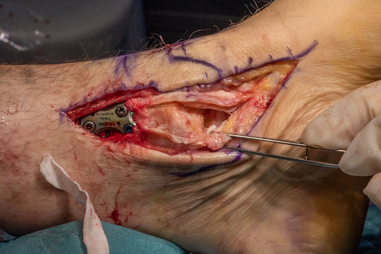
Ankle instability (Medial): Deltoid ligament and tibialis posterior sheath repair.
Mark Herron FRCS
OrthOracle, London, UK.
The indications for surgery to the deltoid ligament can be considered in terms of the acute injury setting, where there is a degree of controversy about the necessity for deltoid ligament repair, and the chronic setting in the presence of longstanding planovalgus deformity and/or medial ankle instability where there is little controversy about repair of the structure. These are discussed in more detail in the indications section.
The deltoid ligament is composed of superficial and deep components which are confluent with each other. The superficial part of the deltoid is the main restraint to ankle and hindfoot valgus and the deep component resists external rotation. Anatomically, the superficial deltoid runs from the medial malleolus as a fan-shaped structure its’ described components being the tibionavicular, tibiospring, tibiocalcaneal and plantar calcaneonavicular. The deep component of the deltoid also takes origin from the tip of the medial malleolus, though inserts in two bands, both into the talus, the anterior tibiotalar and the posterior tibiotalar.
Medial deltoid ligament reconstruction is notorious for its difficulty primarily due to its complex anatomical structure and the difficulties in being able to tension the ligament correctly. Re-ruptures in chronic cases are not infrequent because, suturing of degenerate ligament is difficult, as is the ability to securely attach a thickened and myxoid structure back on to the medial malleolus.
Readers should also familiarise themselves with Kartik Hariharans’ excellent technique :
This contains a very detailed series of images and instructional steps explaining reconstruction of the deltoid ligament using a bone tunnel technique with the Arthrex internal brace implant.
Readers will also find the following associated techniques of interest :
Pes Planus correction with FDL transfer, Calcaneal osteotomy and Wright Bioarch arthroresis screw
Tibialis Posterior reconstruction for pes planus, using FDL transfer and Arthrex Biotenodesis screw.
Lateral ankle ligament reconstruction: Broström (mid-substance) repair
Lateral ankle ligament reconstruction: Brostrom technique