Übersetzt aus dem Englischen:
Deformitäten der langen Knochen in der unteren Extremität können durch Trauma entstehen, sekundär durch Stoffwechselknochenerkrankungen erworben werden oder das Ergebnis angeborener Deformitäten sein. Deformitäten der langen Knochen, die zu einer Abweichung der mechanischen Achse führen, können langfristig zu arthritischen Veränderungen sowohl im Knie als auch im Knöchel führen, wenn sie unbehandelt bleiben. Die Größenordnung der Deformität, die langfristige Gelenkschäden verursachen wird, ist jedoch umstritten und es gibt keine allgemein anerkannten Zahlen. Wahrscheinlich werden Deformitäten bis zu 5 Grad in der koronalen Ebene und 10 Grad in der sagittalen Ebene toleriert, jedoch wird dies von der Lage der Deformität im betroffenen Knochen abhängen.
Bei der Überlegung zur Deformitätenkorrektur ist eine sorgfältige Planung erforderlich. Dies wurde im Fall “Einführung in das Taylor Spatial Frame und Deformitätenplanung” erläutert. Der erste Schritt besteht darin, die mechanische Achse der Extremität vom Zentrum der Hüfte zum Zentrum des Knies zu zeichnen, um festzustellen, ob eine Varus- oder Valgus-Deformität des Beins vorliegt. Als Nächstes identifiziert man, welches Segment der Extremität betroffen ist, indem man die Gelenkwinkel um das Knie zeichnet und diese entweder mit der normalen kontralateralen Seite vergleicht, wenn keine Deformität vorliegt, oder normale Werte aus der veröffentlichten Population verwendet. Dies dient dazu festzustellen, ob die Deformität in der Tibia oder dem Femur oder beiden Knochen liegt. Sobald der betroffene Knochen identifiziert wurde, lokalisiert man die Position der Deformität im Knochen, indem man die Achse der proximalen und distalen Segmente des Knochens zeichnet, um den CORA (Zentrum der Rotationsverformung) zu identifizieren. Mit diesen Informationen sind wir nun in der Lage, die Größenordnung der Deformität zu bewerten, zu planen, wo eine Osteotomie durchgeführt werden soll, um die Deformität zu korrigieren, und zusätzlich zu planen, wo eine physikalische oder virtuelle Gelenkachse platziert werden soll, um die Korrektur durchzuführen, und dann zu entscheiden, wie die Osteotomie während und nach der Korrektur stabilisiert werden soll.
Die Deformitätenkorrektur kann akut erfolgen, wenn die Weichteile dies zulassen, oder allmählich, in welchem Fall den Weichteilen Zeit gegeben wird, sich anzupassen und sich ebenfalls zu korrigieren. Wenn die Deformität akut korrigiert wird, kann die Stabilisierung durch interne Fixierung mittels eines Zwischenaggregatsnagels oder einer Platte angemessen sein. In der Mehrzahl der Fälle einer allmählichen Korrektur ist ein Ringfixateur die bevorzugte Methode der Stabilisierung. Ein weiterer Vorteil der Verwendung eines Ringfixateurs besteht darin, dass er eine Feinabstimmung der Korrektur nach der Operation ermöglicht und eine genauere endgültige Korrektur als bei akuter Korrektur und interner Fixierung ermöglicht.
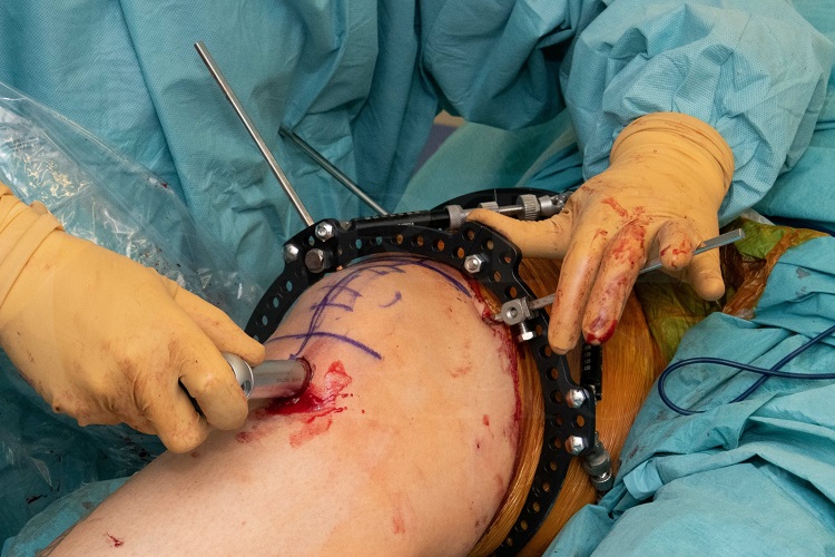
Aufgrund der Herausforderungen bei der akuten Korrektur komplexer Deformitäten, insbesondere wenn sie in mehr als einer Ebene vorliegen, haben sich verschiedene Methoden entwickelt, um die Vorteile einer genaueren Korrektur mit einem externen Fixateur zu kombinieren, zusammen mit der Verwendung von interner Fixierung zur Stabilisierung der endgültigen Korrektur und somit die Notwendigkeit für eine längere Zeit in einem externen Fixateur für den Patienten zu vermeiden. Eine solche Methode ist die computergestützte hexapodische orthopädische Chirurgie (CHAOS-Technik), wie sie von der Bristol Limb Reconstruction Unit beschrieben wurde (siehe unten stehende Referenz). Diese Methode verwendet einen Hexapod-Rahmen, um komplexe Deformitätskorrekturen akut im Operationssaal durchzuführen. Nach Abschluss der Korrektur wird die Osteotomie mit interner Fixierung stabilisiert und der Rahmen wird dann entfernt. Diese Technik ermöglicht eine genaue Deformitätskorrektur, ohne dass eine längere postoperative Zeit in einem Ringfixateur erforderlich ist.
Original Intro:
Femoral deformity correction: CHAOS technique using Taylor Spatial Frame and Trigen Nail (Smith and Nephew)
Mr Paul Fenton FRCS (Tr & Orth)
The Queen Elizabeth Hospital, Birmingham, UK.
Long bone deformity in the lower limb may follow trauma, be acquired secondary to metabolic bone disease or may be the result of congenital deformities. Deformity in the long bones which results in a mechanical axis deviation can, in the longer term, result in arthritic changes in both the knee and the ankle if untreated. The magnitude of deformity which will result in longer term joint damage is however controversial with no universally accepted figures. Probably deformities up to 5 degrees in the coronal plane and 10 degrees in the sagittal plane are tolerated, however this will depend on the location of the deformity in the affected bone.
When deformity correction is considered careful planning is required. This has been outlined in the ‘Introduction to the taylor spatial frame and deformity planning’ case. The first step involves plotting the mechanical axis of the limb from the centre of the hip to the centre of the knee to determine whether a varus or valgus deformity of the leg is present. Next, one identifies which segment of the limb is involved by plotting the joint line angles around the knee and comparing these either with the normal contralateral side if no to deformity is present theret or using published population normal values. This one to identify whether the deformity lies within the tibia or the femur or both bones. Once the involved bone has been identified one locates the position of the deformity in the bone by plotting the axis of the proximal and distal segments of the bone to identify the CORA (centre of rotation of angulation). With this information we are now able to assess the magnitude of the deformity, to plan where to perform an osteotomy to allow the deformity to correct and in addition plan where to place either a physical or virtual hinge to perform the correction and then to decide how to stabilise the osteotomy during and after correction.
Deformity correction can be performed acutely, when the soft tissues will allow this, or gradually in which case the soft tissues have time to adapt and correct also. If the deformity is corrected acutely then stabilisation with internal fixation using either an intermediary nail or plate may be appropriate. In the majority of cases of gradual correction a ring fixator is the preferred method of stabilisation. A further benefit of using a ring fixator is that it will allow fine tuning of the correction post-operatively and a more accurate final correction then with acute correction and internal fixation.
Due to the challenges of acutely correcting complex deformities, particularly when in more than one plane, using standard internal fixation techniques various methods have developed to combine the benefits of more accurate correction with external fixator together with the use of internal fixation to stabilise the final correction and thereby avoiding the need for prolonged time in an external fixator for the patient. One such method is the computer hexapod assisted orthopaedic surgery (CHAOS) technique as described by the Bristol Limb Reconstruction unit (see reference below). This method uses a hexapod frame to perform complex deformity correction acutely in theatre. Once the correction has been completed the osteotomy and is stabilised with internal fixation and the frame is then removed. This technique allows accurate deformity correction without the need for prolonged postoperative time in a ring fixator.
Hughes A, Heidari N, Mitchell S, Livingstone J, Jackson M, Atkins R, Monsell F. Computer hexapod-assisted orthopaedic surgery provides a predictable and safe method of femoral deformity correction. Bone Joint J . 2017 Feb;99-B(2):283-288.
Readers will also find the following associated OrthOracle techniques of interest:
Tibial fracture non-union correction using Taylor Spatial Frame (Smith and Nephew)
Supra-malleolar distal tibial osteotomy: Minimally invasive technique with the Taylor Spatial Frame